Area measurement methods are used in various clinical practices such as wound management, dermatology, plastic and reconstructive surgery, burn units, surgical specialities and more. Traditional methods are often time-consuming or inaccurate. With this brief overview of existing methods, you will learn how imito solutions provide a safe, effective and fast alternative, directly on your smartphone.
Overview of Area Measurement Methods
Area measurement methods are mainly but not exclusively used in wound care to objectively document the lesion evolution; Is it growing bigger? How far does it extend? How fast is the size evolving? Does it shrink as expected in response to a specific treatment? For that different methods can be used and based on the excellent systematic review by Jørgensen LB et al. [1], we summarised the pros and cons for each of the two-dimensional measurement methods.
Methods involving skin contact
- Planimetric method: After having traced the wound border on a metric gridded layer (scale paper), the number of small squares is counted and returns the surface area. This manual method is accurate and reliable but takes time and involves skin contact and therefore a contamination risk.
Methods without skin contact
- Simple ruler method: The length and width are multiplied to estimate the area. This method is easy and low cost, but very often highly overestimates the surface, sometimes up to 40 %!
- Mathematical models: The elliptical method or the Kundin wound gauge (length × width × 0.785) are used to estimate area using a mathematical formula. Those methods are easy to use and the surface is quickly calculated. Their assumption of an elliptical/rounded lesion shape makes them unfortunately inaccurate for irregular wounds.
- Stereophotogrammetry: A special stereographical camera (taking several pictures with different angles) supported by a computer can return the wound area, length and width. This method is accurate but time-consuming, expensive, and requires the specific camera to be available at the bedside.
- Digital imaging: An object (e.g. a ruler) with a known size is placed near the wound. The first step, after taking a picture, is to select the object or a part of it to serve as a reference size. In the second step, the border of the wound is traced and calculated. This method is accurate and as reliable as planimetry, but its two steps make it time-consuming.
imito’s Method: Digital Planimetry
With imito digital planimetry, we are offering healthcare professionals a contactless, accurate, reliable and fast method to measure a surface area and all of this directly on a smartphone!
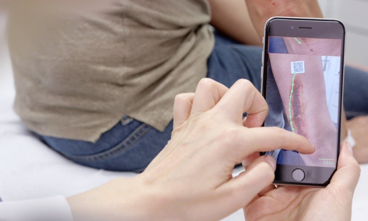
The process is as follows:
- The user places the Calibration Marker (also available as single-use stickers) next to the region of interest.
- The user takes a picture of the region. The image is automatically calibrated.
- The user then uses his/her finger to draw a border of the region of interest directly on his smartphone screen. The app calculates the exact surface of the polygon using digital planimetry.
During our validation tests, we came to the following results:
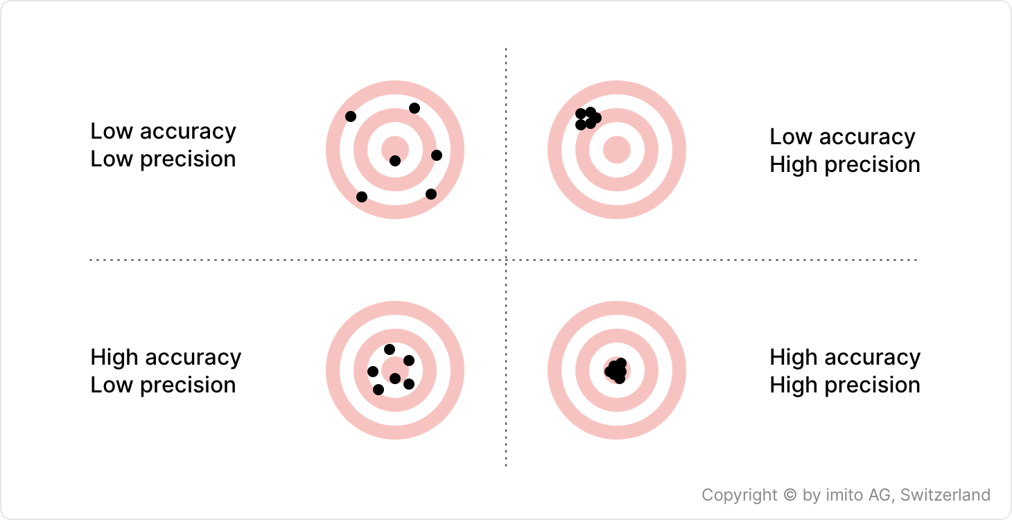
- Accuracy: expressed as an experimental error of about 5 % for width and length measurements and up to 10 % for surface area.
- Precision: expressed as coefficient of variation (% RSD) < 5 %.
Measurement quality depends on random errors such as environment (distance to marker or small tilt of the marker that makes it not exactly parallel to the picture), inter-operator variations (when tracing the wound boundaries on screen) and systematic errors (device camera optics distortion). Research findings consistently highlight the advantages of imito's digital planimetry over conventional methods. According to French clinical researchers, our measurement technology demonstrated “good validity and reproducibility” in the clinical evaluation of pressure ulcers [2].
imito’s measurement method was compared with the ruler and acetate tracing methods, which are currently the reference standards for measuring wounds. The study found that imito's digital planimetry method had “excellent validity” and “intra- and inter-rater reproducibility” for wound length, width, and surface area measurements. Another study has demonstrated that our digital planimetry offers superior accuracy and reliability compared to the ruler method [3].
In contrast to laser-based methods, which often require expensive equipment and technical expertise, imito's digital planimetry presents a cost-effective and user-friendly alternative. A study conducted by researchers at the University Medical Center in the Netherlands showed that our measurement technology exhibited exceptional concurrent validity and reliability in measuring the area of concave wounds, establishing its credibility as a dependable instrument in both clinical research and everyday healthcare practice [4].
Moreover, when compared to digital photography and the ImageJ processing tool, our measurement system was proven to be a useful and practical method for area measurement with excellent repeatability and accuracy [5].
These findings collectively underscore the potential of imito's digital planimetry as a superior tool for wound measurement, offering distinct advantages in terms of precision, consistency, and accessibility.


